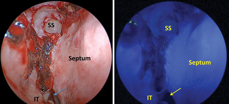
Fig. 1.

True-color and ICG-fluorescence images of the nasoseptal flap just before harvest in the patient with a type-2 craniopharyngioma (patient 1). In this case, the flap enhanced so avidly and uniformly that the enhancement of the underlying arterial supply was not discernable. ICG, indocyanine green; IT, inferior turbinate; SS, sphenoid sinus.
