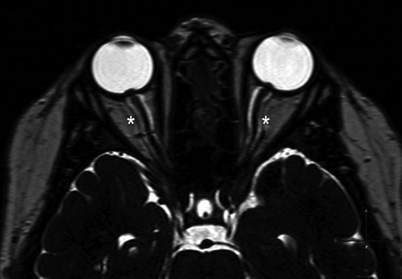
Fig. 3.

Dilation of optic nerve sheaths: MRI scan, axial section, heavily-weighted T2 sequence, shows enlarged CSF spaces appearing as a high-signal intensity around the optic nerves (asterisks). This is the scan of a 76-year-old woman, overweight, who presented CSF leak from the posterior wall of the right sphenoid sinus. CSF, cerebrospinal fluid; MRI, magnetic resonance imaging.
