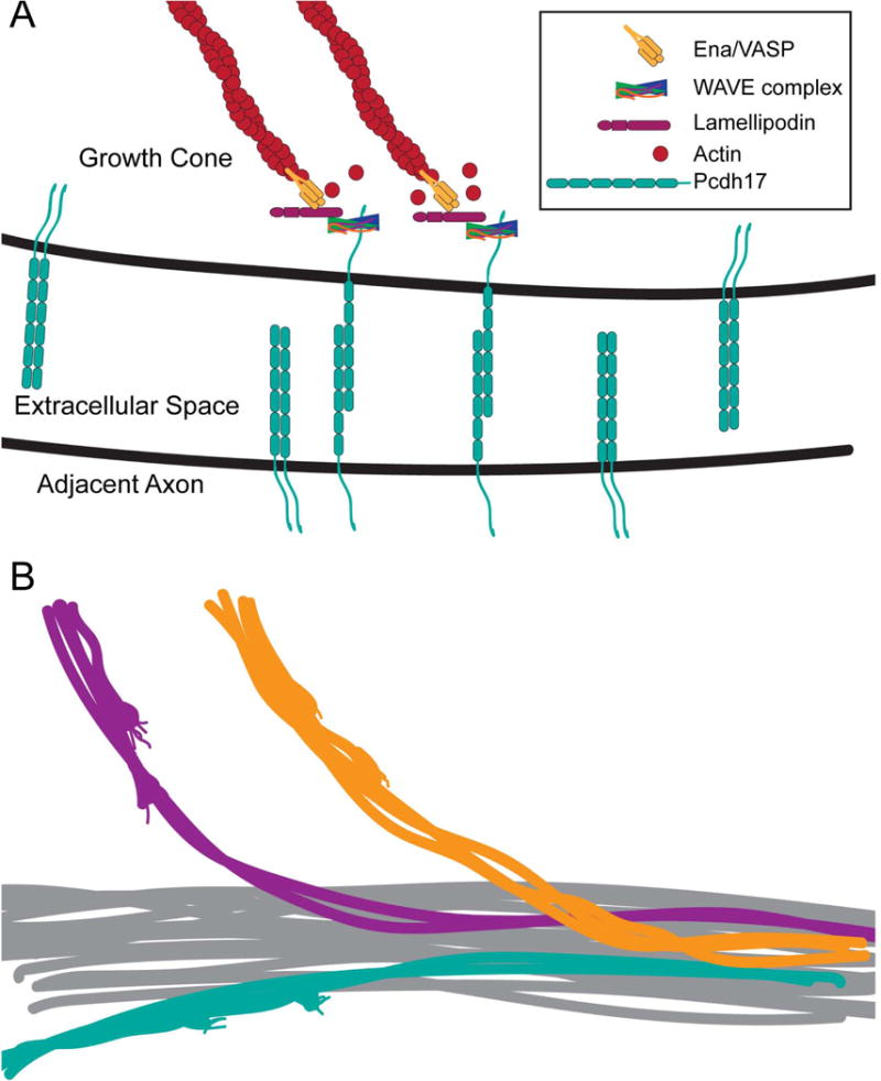
Figure 3. Contact-dependent axon outgrowth mediated by δ2-protocadherins.

A. Model based on Hayashi et al. (2014). Shown here are homophilic interactions between Pcdh17 molecules on the apposing plasma membranes of a migrating growth cone and an axon. Pcdh17 recruits the WAVE complex, which interacts directly with a conserved motif. Pcdh17-WAVE complex recruits the scaffolding protein, Lamellipodin, and Ena/VASP to the site of contact. Ena/VASP promotes the addition of actin monomers to the plus-ends of actin filaments. This contact-dependent growth both enhances and directs motility, so that follower axons that express Pcdh17 move efficiently along Pcdh17+ axons.
B. If each of the δ2-pcdhs, which also have the conserved WAVE binding motif, acts similarly to Pcdh17, this would promote the selective formation of axon bundles on the basis of differential protocadherin expression, as well as segregation of axons withina larger axon tract. Similarly, this could also promote the selective de-fasciculation at exit points. Promoting selective axon bundling would increase the robustness of axon targeting. If active at early stages of axon guidance, this could also increase the reliability of path finding, as clusters of growth cones would collaborate to respond to cues, rather than individual pioneers.
