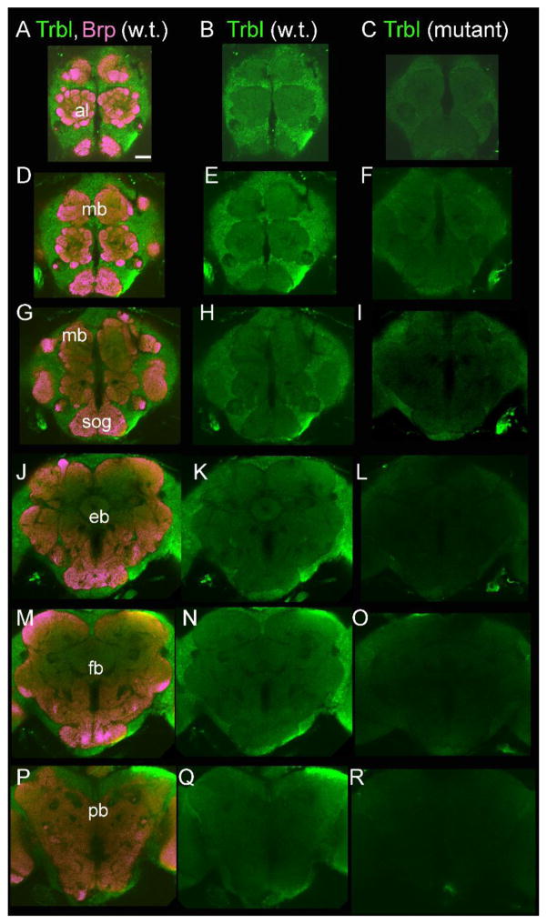
Figure 3. Tribbles immunostaining in the Drosophila brain.
Trbl is expressed in cell bodies and the neuropil throughout the fly brain visualized in green in both wild-type CS brains (left and center columns) and trbl mutant fly brains (right column). Mutant flies have reduced Tribbles expression compared to wild-type CS levels, comparing center and right most panels. Brains were co-labeled with the bruchpilot (brp) monoclonal antibody marking the synapses in purple on the left most panels. Labeled structures: antennal lobes (al), mushroom bodies (mb), subesophogial ganglia (sog), ellipsoid body (eb), fan shaped body (fb), protocerebral bridge (pb). Scale bar = 50 μm.

