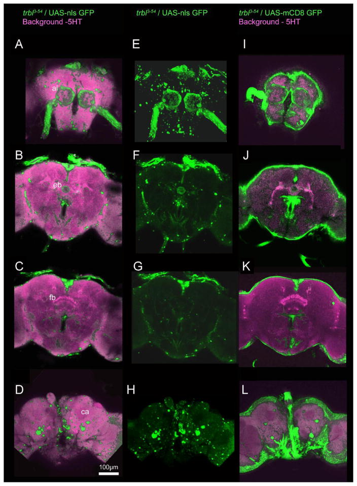
Figure 5. trbl3-54 Gal4 driven expression of two different UAS-GFPs in the adult brain.
GFP expression is visualized from anterior to posterior A–D with UAS-nls-GFP and anti-5HT, E–H with UAS-nls-GFP alone, and I–L with UAS-mCD8-GFP and anti-5HT. Notable structures are labeled: antennal lobes (al), median bundle (meb), ellipsoid body (eb), fan shaped body (fb) and calyces (ca). Expression is visualized throughout the brain including in the antennal lobes, median bundle, ellipsoid body, and around the periphery of the brain. Scale bar = 100 μm.

