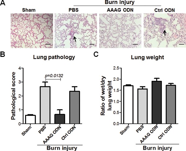
Figure 5.

Alleviating role of AAAG ODN on ALI of burn-injured mice. The lungs of the mice at 24 h post burn were weighed and sectioned for H&E staining. The slices were (A) observed for pathological changes and (B) evaluated for pathological scores. (C) The wet/dry weight ratio of lung was used as a parameter to denote acute pulmonary edema. The black arrows indicate the infiltrated inflammatory cells. Data are represented as mean ± SEM (n = 3 mice/group). *P < 0.05, **P < 0.01, ***P < 0.001. Scale bars = 50 μm, magnification x 200.
