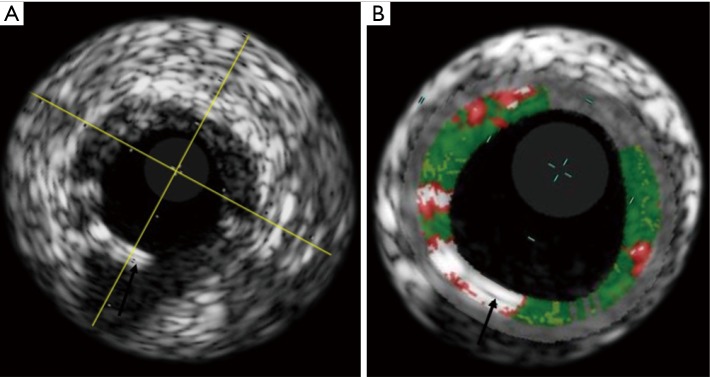
Figure 3.
(A) Shows a greyscale IVUS image of an Absorb BVS after implantation. In (B), the VH is superimposed to the greyscale image, showing how the Absorb BVS struts (black arrows) are recognized by the software as dense calcium (white) and necrotic core (red). IVUS, intravascular ultrasound, VH, virtual histology.

