Figure 9.


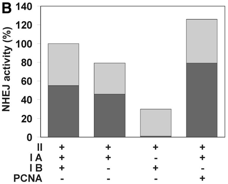
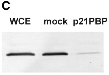
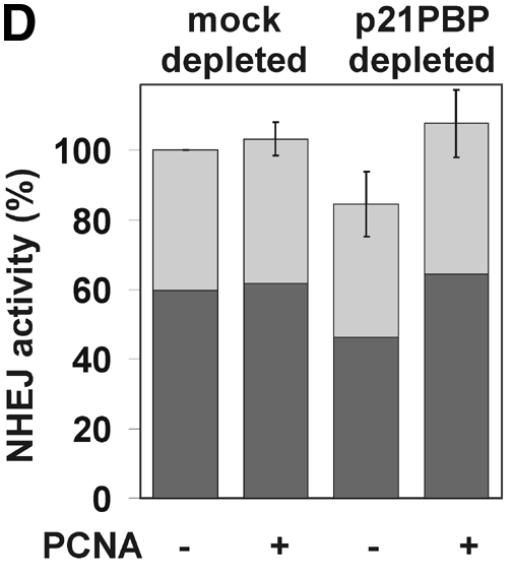
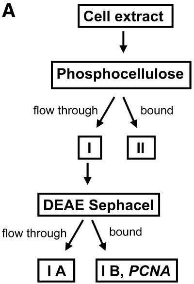
The effect of PCNA on NHEJ. (A) Flow diagram of fractionation of the HeLa cell extract to separate PCNA. As described in Materials and Methods, HeLa whole cell extract was passed through a phosphocellulose column, resulting in fractions I (flow-through) and II (bound). PCNA-containing fraction I was further separated through DEAE–Sephacel to generate fractions IA and IB, the latter containing PCNA. (B) DSB substrate XM was incubated with combinations of different fractions from the above fractionation scheme as indicated. Substrate DNA was recovered to assess formation of circular monomeric end joining products as described in Materials and Methods, defining the NHEJ activity of the combined fractions IA, IB and II as 100%. The fraction of blue plaques is indicated in black and the fraction of white plaques in gray. Note that substitution of fraction IB with purified PCNA restores NHEJ to >100%. (C) Western analysis indicating the removal of PCNA from HeLa cell extract. PCNA was depleted from HeLa whole cell extract with a p21-derived peptide (PBP for PCNA-binding peptide) or mock-depleted with a mutated p21-derived peptide (mock). The mutated p21-derived peptide was incapable of precipitating PCNA and served as a negative control. PCNA was detected in cell extracts by western analysis as described in Materials and Methods. (D) Depletion of PCNA causes a small but significant reduction in NHEJ activity in HeLa cell extracts. NHEJ activity of PCNA-depleted and mock-depleted HeLa cell extract was determined as described in Materials and Methods. NHEJ activity of the mock-depleted cell extract was arbitrarily taken as 100%. Error bars indicate the standard deviation of total NHEJ activity. The reduction in NHEJ in PCNA-depleted cell extracts was highly significant in eight independent experiments (P = 0.0012, one-sided Student’s t-test).

