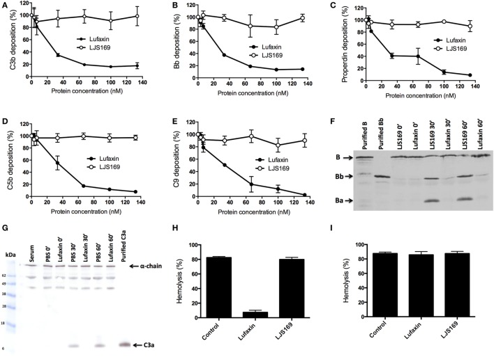
Figure 3.
Lufaxin acts exclusively at the early steps of the alternative pathway (AP) of the complement system. (A–E) Normal human serum (NHS) was added to agarose-coated microplates together with Lufaxin or LJS169 (as negative control) and incubated at 37°C for AP activation. Deposition of C3b (A), Bb (B), properdin (C), C5b (D), and C9 (E) was assessed using specific antibodies. (F,G) AP-mediated hemolysis assays were performed in the presence of Lufaxin, LJS169 or PBS. At different times of incubation (0′, 30′, and 60′), supernatants were collected and submitted to SDS-PAGE. Proteins were transferred to nitrocellulose membranes and then incubated with anti-factor B (F) or anti-C3a antibodies (G). A control aliquot with diluted NHS without inhibitor and red blood cells (Serum) was also loaded onto the gel. (H,I) Effect of Lufaxin or LJS169 on formation of the membrane attack complex. Rabbit erythrocytes were first incubated with C6-depleted serum at 37°C, and after incubation, the cells were centrifuged and the supernatant discarded. The cells were then resuspended with NHS in EDTA buffer (blocking the initial steps of the cascade) and incubated again. Lufaxin or LJS169 was added in the first (H) or second (I) incubation.

