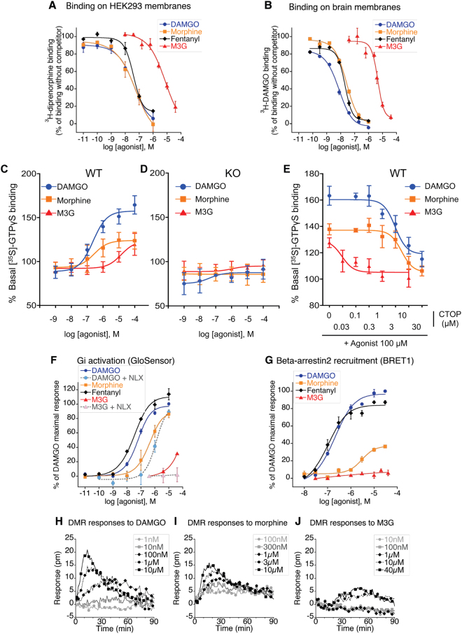
Figure 6.
M3G binding and signaling to MOR (A,B) [3H]-diprenorphine binding on membranes from MOR expressing HEK293-Glo cells (A) and [3H]-DAMGO binding on brain membranes preparations from WT mice (B). Membranes were incubated with increasing doses of DAMGO, morphine, fentanyl or M3G in assay buffer containing a fixed dose of opioid radioligand. 100% represents maximal radioligand binding in the absence of competitor. Results are presented as means ± SEM of 2 or 3 experiments. (C–E) MOR agonist-induced [35S]-GTPγS binding to brain membranes preparations from WT (C) or KO mice (D). Membranes were incubated with increasing doses of DAMGO, morphine or M3G agonists (10−9 to 10−4M) in assay buffer containing [35S]-GTPγS. Basal level (100%) represents [35S]-GTPγS binding in the absence of agonist. DAMGO, morphine and M3G significantly stimulated [35S]-GTPγS binding to membranes from WT but not KO mice. Results are presented as means ± SEM of 3–5 experiments on 3 independent membrane preparations per genotype. (E) The selective MOR antagonist CTOP inhibits DAMGO, morphine and M3G-induced [35S]-GTPγS binding to membranes from WT mice. Brain membranes from WT mice were incubated with increasing doses of the mu opioid antagonist CTOP combined with 100uM of DAMGO, morphine or M3G. Activation results are presented as means ± SEM of 6–10 experiments from 3 independent membrane preparations. (F) Effect of increasing concentrations of DAMGO, morphine, fentanyl and M3G on forskolin-stimulated cAMP production in HEK293 stably expressing GloSensor and MOR receptor. In the cases indicated, 1 µM of naloxone had been added to the cells 15 min prior to the agonist. Dose−response curves were normalised to maximal DAMGO activity. Evaluations were performed three times in duplicate for M3G, two times in duplicates for other agonists. Results are presented as means ± SEM. (G) eYFP-labelled Beta-arrestin-2 translocation to Rluc-MOR in HEK293 cells after 5 to 10 min of cells activation by DAMGO, morphine, fentanyl or M3G at 37 °C. Agonist specific BRET1 ratio were determined by subtracting BRET1 ratio of non activated cells, and normalised to maximal DAMGO-triggered effect. Presented results are means ± SEM of 2 to 4 experiments. (H–J) Dynamic mass redistribution (DMR) signals observed in HEK293-Glo-cells after activation by various concentrations of DAMGO, morphine or M3G. Baseline of buffer-treated cells has been subtracted. Evaluations were performed three times in duplicate or triplicates. Presented figures show a representative experiment.

