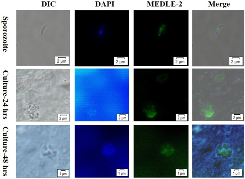
FIGURE 3.

Localization of MEDLE-2 protein expression on Cryptosporidium parvum sporozoites (Top) and developmental stages of C. parvum in HCT-8 cell cultures at 24 and 48 h (Middle, Bottom, respectively). Images were taken by differential interference contrast microscopy (DIC), fluorescence microscopy using nuclear stain 4, 6-diamidino-2-phenylindole (DAPI), fluorescence microscopy using polyclonal MEDLE-2 antibodies (MEDLE-2), and superimposition of the three (Merge).
