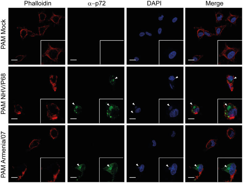
Figure 7.
Viral factory pattern in PAM. Cells were infected with NHV/P68 and Armenia/07 strains (MOI = 1), fixed at 16hpi and incubated with phalloidin-TRITC, anti-p72 antibody and DAPI to respectively stain actin filaments, viral particles and cellular and viral DNA. Z-slides images were taken by CLSM and represented as a maximum of z-projection. Arrowheads show the viral factories visualized by anti-p72 MAb and with DAPI to stain the viral DNA. Images are representative of two independent experiments.

