
Figure 6.
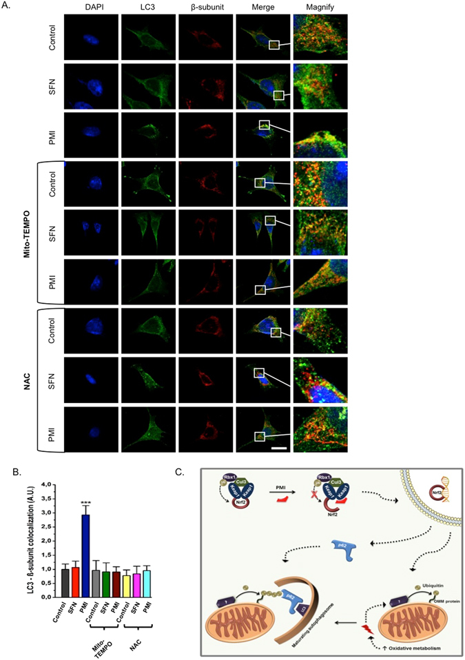
Selective scavenging of mitochondrial superoxides inhibits the PMI-mediated initiation of mitophagy. (A) Representative high-resolution images of LC3 localization in MEF cells treated with DMSO vehicle control, 1 μM SFN, or 10 μM PMI for 24 h in the presence or absence of 50 µM mito-TEMPO or 5 mM NAC. Cells were immunolabeled for LC3 (green) and β-subunit (red). Scale bar represents 10 μm. A magnification of the merge images is shown in areas demarcated by the white box. (B) Quantification of the extent of LC3:β-subunit co-localization (n > 10, ***p < 0.001). (C) Inhibition of the PPI between Keap1 and Nrf2 leads to the nuclear accumulation of the latter and the subsequent transcriptional activation of genes involved in the regulation of mitochondrial function and autophagy, including p62. As a result, the rate of oxidative metabolism is increased and the mitochondrial redox status is altered, which initiates a mitophagic response that targets mitochondria to autophagosomes via p62.