
Fig. 5.
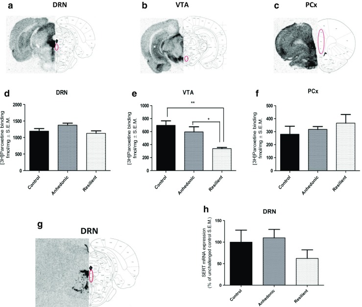
Representative autoradiorgams of specific [3H]paroxetine binding to SERT protein in DRN (a), VTA (b), and PCx (c) and SERT mRNA (g) in coronal sections of the rat brain. Brain structures were dissected according to Paxinos and Watson (1998). The effect of stress response on SERT level was investigated in DRN (d), VTA (e), and PCx (f). h SERT mRNA expression in DRN. One-way ANOVA analysis followed by Tukey’s post hoc test showed significant decrease in [3H]paroxetine binding to SERT protein in VTA of resilient animals as compared to control and anhedonic rats (e). n = 4–6; *p < 0.05 vs anhedonic, **p < 0.01 vs control