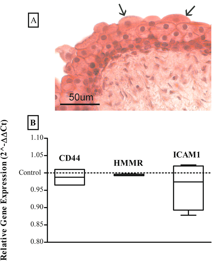
Figure 8.

Light microphotograph (A) with haematoxylin and eosin (HE) stain showing histologic evaluation of rabbit bladder (Top to base: umbrella, intermediate and basal cells; arrows show the typical “dome-shaped” apical cells). Normalized expression value 2−ΔΔCt (mean ± standard error) (B) of CD44, RHAMM and ICAM-1 in bladder tissues incubated with HA-AuNP and CS-AuNP (left box for each type of receptor) and control bladder tissues incubated with BSA-AuNPs solution (dashed line).
