Figure 1.
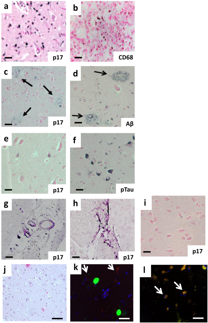
Human HIV-positive brains showed p17-positive staining in inflammatory and neurodegenerative regions. Representative immunohistochemistry of brain sections showing the staining for (a) p17 (N-DAB stain, gray-black) and (b) CD68-positive macrophages localized to the same region in serial sections as did (DAB stain, brown), (c) p17 (N-DAB) and (d) β-amyloid (Aβ, DAB stain) positive plaques (black arrows). (e) P17-positive (N-DAB) and (f) phosphorylated tau (p-tau, DAB stain) positive cortical neurons from the same region. (g) Medium sized cortical blood vessels were positive for p17 and (h) p17-positive fibril structures within the cortex. (i) Shows negative expression of p17 in the cortical region from a non-HIV positive individual and (j) a negative control, where the p-17 primary antibody was replaced with PBS during immunohistochemical staining. (k) Representative immunofluorescent co-localization of p17 (TRITC-red) and p-tau (FITC-green) and (l) β-amyloid (Aβ, FITC-green) in adjacent neurons within the cortex. Nuclei were counter-stained with DAPI (blue). A similar pattern for p17 where yellow/orange demarcates overlapping staining (white arrows) was evident. Representative images are shown from three patient tissue samples used in this immunohistochemical study.(a–j) Scale bar = 50 µm and (k,l) = 20 µm.

