Figure 7.
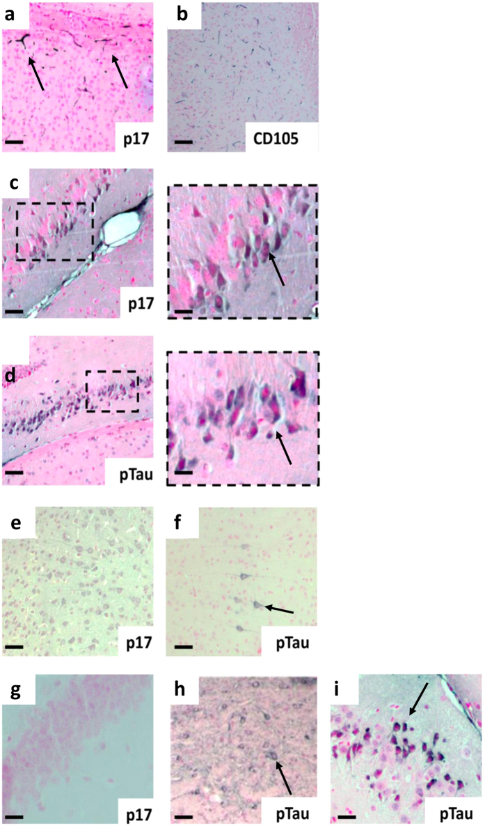
Histological localization of p17 in the mouse hippocampus and local cortical regions. Representative immunohistochemistry of brain sections showing the staining for (a) p17 (N-DAB stain, gray-black) into the CA1 region localized to cortical microvessels and (b) CD105 (DAB)-positively stained vessels in the same location in a serial section. (c) P17 was observed in hippocampal neurons adjacent to the injection point (inset scale bar = 10 µm), (d) in the same region as phosphorylated tau (p-tau, DAB) (inset scale bar = 10 µm). (e) P17 was found to be expressed consistently within cortical neurons, some of which (f) were also p-tau-positive. (g) No staining was observed in none-injected controls. (h,i) P-tau positivity in sham-injected 3xTg mouse. Scale bars (a–h) = 50 µm, (i) = 10 µm. Images show representative staining results from one or more of six animals in each grouping where sectioning throughout the whole bregma was carried out. See Supplementary Information for immunofluorecence and double immunohistochemistry staining patterns as well as primary antibody-negative controls.

