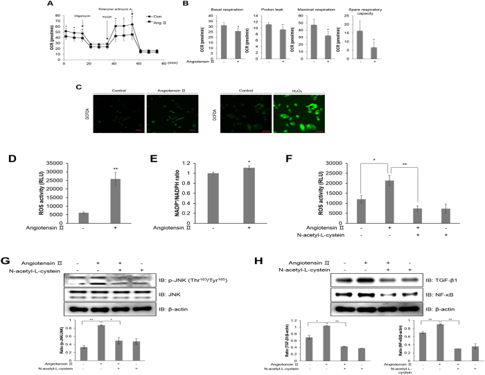
Figure 2.
Ang ІІ regulates mitochondrial oxygen consumption and generates ROS in HL-1 atrial cells. (A) Mitochondrial oxygen consumption rate (OCR) in HL-1 cells was measured using the Seahorse flux analyzer in response to 1 μM oligomycin, 1 μM FCCP, and 0.5 μM rotenone/antimycin (A). (B) Basal respiration, proton leak, maximal respiration, and spare respiratory capacity were calculated from (A). (C) HL-1 cells were pre-treated with 10 μM DCFDA for 40 min, and then treated with 1 μM Ang ІІ and 100 μM H2O2, respectively. H2O2 was used as a positive control. ROS levels were assessed using a confocal microscope in real-time. (D) HL-1 cells were treated with 1 μM Ang ІІ for 24 h. The cells were lysed with ROS detection solution, and the concentration of ROS was evaluated by colorimetric absorbance assay. (E) HL-1 cells were stimulated with 1 μM Ang ІІ for 24 h, and the NADP+/NADPH ratio was calculated by NADP+/NADPH colorimetric assay. HL-1 cells were pre-treated with 10 mM N-acetyl-L-cysteine (NAC) for 30 min and then treated with 1 μM Ang ІІ for 24 h. (F) ROS levels as measured by a colorimetric absorbance assay. (G) JNK phosphorylation was quantified by western blot analysis using antibodies specific to the phosphorylated protein. Levels of total JNK and β-actin were also measured as protein loading controls. (H) TGF-β1 and NF-κB protein levels were assessed by immunoblotting with specific antibodies. *P < 0.05, **P < 0.01, compared with the untreated cells. Results are from three independent experiments.

