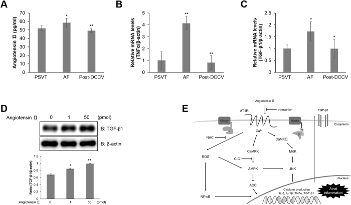
Figure 7.
Ang ІІ is increased in patients with atrial fibrillation. (A) The concentration of Ang ІІ in plasma samples from patients with PSVT (n = 6), DCCV (n = 11), and post-DCCV (n = 11). (B) Total mRNA was extracted from 50 μL of plasma samples from patients with PSVT (n = 6), DCCV (n = 11), and post-DCCV (n = 11), and quantitative RT-PCR analysis was performed with specific human primers targeted to TNFα and β-actin. β-actin was used as an endogenous control. (C) Total mRNA was extracted from 50 μL of plasma samples from patients with PSVT (n = 6), DCCV (n = 11), and post-DCCV (n = 11), and quantitative RT-PCR analysis was performed with specific human primers targeted to TGF-β1 and β-actin. β-actin was used as an endogenous control. (D) HL-1 cells were stimulated the indicated doses of Ang ІІ for 24 h. Cell lysates were analysed by western blot analysis using with antibody against TGF-β1. Levels of β-actin was used as control. (E) Schematic representation of the signalling pathway underlying atrial fibrillation. *P < 0.05, **P < 0.01 compared with the untreated cells. Results are from three independent experiments.

