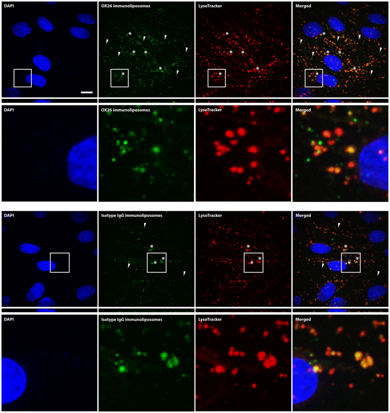
Figure 3.
Spinning disk confocal microscopy images of immunoliposome-treated, primary rat BCECs. Treatment with either OX26 (upper panel) or isotype IgG (lower panel) immunoliposome revealed a clear association between the fluorescently labelled immunoliposomes and the BCECs. Morphologically, the fluorescent signals were either particulate in the periphery of the cells (arrows) or clustered into larger structures in the perinuclear area (asterisks). Counterstaining with LysoTracker revealed that these larger structures were lysosomes that the endocytosed immunoliposomes had been sorted to. The smaller, particulate signal (arrows) did not co-localize with the lysosomes, suggesting these to be newly endocytosed immunoliposomes. Scale bar depicts 10 µm. DAPI: Diamino-phenylindole.

