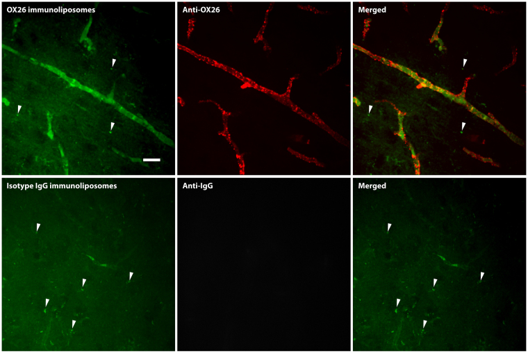
Figure 5.
Uptake of fluorescently labelled immunoliposomes in brain capillaries in vivo evaluated by spinning disk confocal microscopy. Young rats were injected with either OX26 (upper panel) or isotype IgG (lower panel) immunoliposomes to assess their potential of interacting with the capillaries of the brain. OX26 immunoliposomes (upper panel) showed a good association to the microvessel structures of the brain, although the fluorescent intensity of the immunoliposomes were very low. Counterstaining against the OX26 antibody revealed that the immunoliposome and ligand had accumulated in the brain capillaries, but no sign of transcytosis could be detected. Isotype IgG immunoliposomes (lower panel) had almost no association to the microvessel structures of the brain, which was also evident from the counterstaining against mouse IgG, which did not present with the same vessel pattern as the OX26 immunoliposomes. In both cases, artefacts from the paraformaldehyde fixation could easily be detected (arrows), which was not regarded as a transcytosed immunoliposome, since the same was observable in non-treated animals (data not shown). Scale bar depicts 20 µm.

