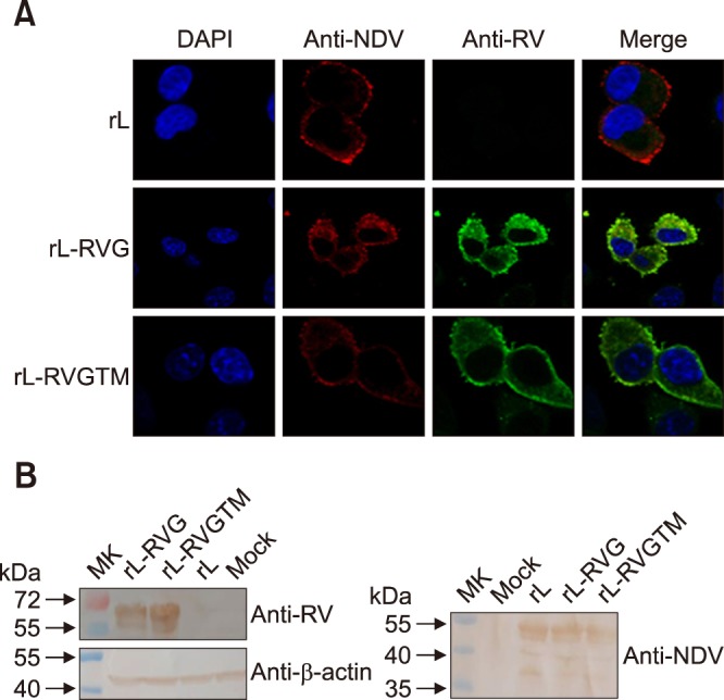
Fig. 2. The rL-RVGTM expresses RV and NDV components. (A) Detection of RVGTM expression by immunofluorescence. BHK-21 cells were infected with rL, rL-RVG, or rL-RVGTM at a multiplicity of infection of 0.1. At 24 h post-infection, the cells were fixed and stained with chicken anti-NDV and dog anti-RV sera followed by fluorescein isothiocyanate (FITC)-conjugated rabbit anti-dog and tetramethyl rhodamine isocyanate (TRITC)-labeled goat anti-chicken antibodies. The cells were permeabilized with 0.02% Triton X-100 and cell nuclei were stained with DAPI. (B) Western blot analysis of recombinant NDV expressing RVGTM. Lysates of BHK-21 cells infected with rL, rL-RVG, or rL-RVGTM were separated by 12% SDS-PAGE and incubated with dog serum against RV, chicken serum against NDV, or anti-β-actin monoclonal antibody as a loading control. After incubation with peroxidase-conjugated secondary antibody, the proteins were visualized with 3,3-diaminobenzidine reagent. The locations of marker proteins are indicated on the left, and the antiserum or antibody used is indicated on the right. RVGTM, chimeric rabies virus G protein; RV, rabies virus; NDV, Newcastle disease virus; RVG, rabies virus G protein; MK, protein molecular marker.

