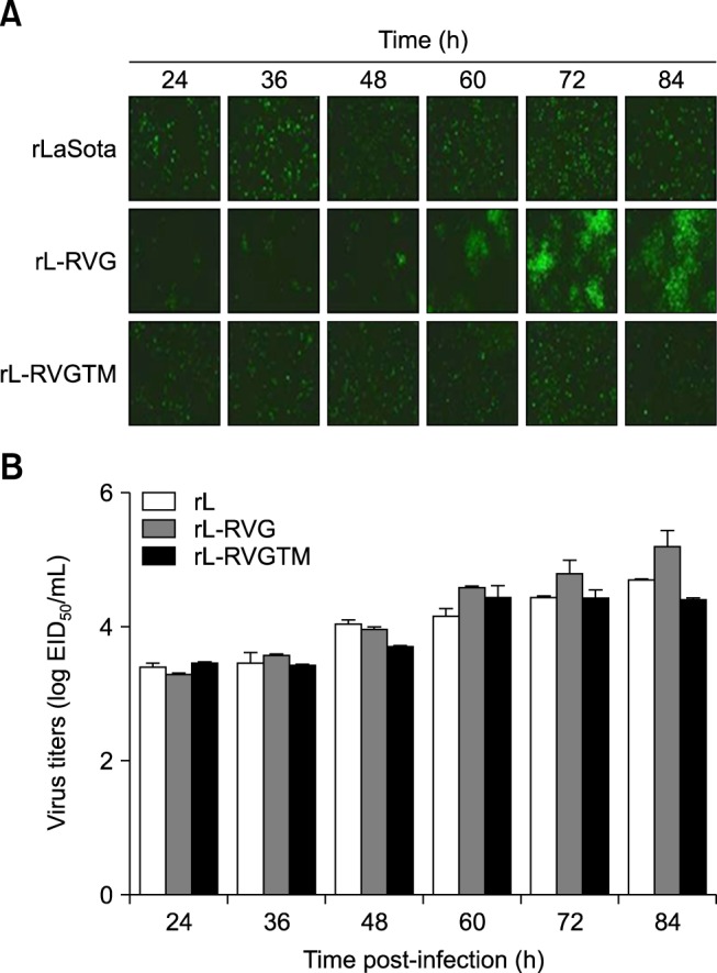
Fig. 5. rL-RVGTM shows a pattern of spread that is similar to the pattern of rL. (A) Immunofluorescence was used to examine the spread of the viruses. BHK-21 cells were infected with rL, rL-RVG, or rL-RVGTM for 1 h at a multiplicity of infection of 0.1, followed by washing in phosphate belanced solution (PBS) and incubation at 37℃. The infected cells were fixed at the indicated times (24, 36, 48, 60, 72, or 84 h) for immunofluorescent staining with chicken serum against NDV. As shown, rL-RVGTM lost the ability to spread from the initial infected cell to adjacent cells. (B) Supernatants were collected at the same times as in panel A to measure virus titers of 10-day-old embryonated chicken eggs. As shown, the peak titer of rL-RVGTM was similar to that of rL but was approximately one-fifth of a log lower than that of rL-RVG. RVGTM, chimeric rabies virus G protein; RVG, rabies virus G protein.

