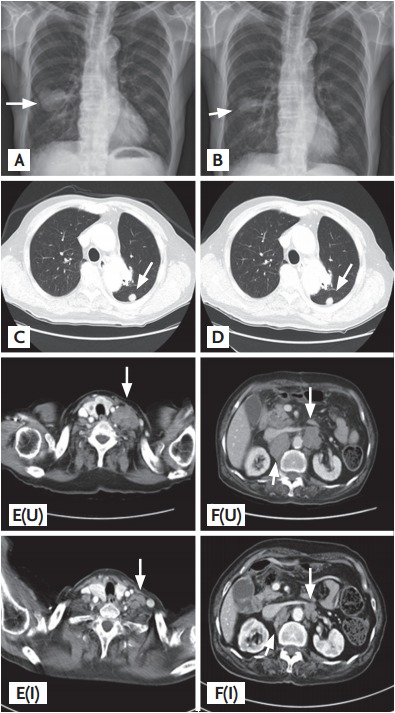
Figure 1.

(A, B) Chest posteroanterior shows 5 cm-sized irregular mass (arrows) on right middle lobe (A). The mass much regressed after 3 months of capecitabine treatment (B). (C, D) Chest computed tomography (CT) shows no remarkable change of metastatic nodule (arrows) in left lower lobe superior segment. (E, F) Chest CT shows enlarged metastatic nodule (arrow) in left supraclavicular region (upper panel of E) and abdomen CT shows multiple metastatic lymphadenopathy (arrow) in portocaval, left para-aortic and aortocaval area (lower panel of E). After 3 months of capecitabine treatment, metastatic lymphadenopathy of the same lesion was much regressed (F).
