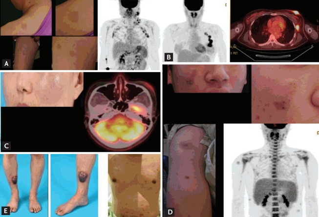
Figure 1.
Initial presentation of patients. (A) Patient (#8) presented with multiple skin lesions and lymph node enlargement on positron emission tomography/computed tomography. (B) Patient (#2) displayed breast skin lesions with increased fluorodeoxyglucose (FDG) uptake and axillary lymph node enlargement. (C) Patient (#6) showed a localized skin lesion of the cheek with moderate FDG uptake. (D) Patient (#7) presented with multiple skin lesions and hepatosplenomegaly. (E) Patient (#9) initially showed a solitary skin lesion of the lower leg (left); however, multiple lesions developed on the trunk, indicating relapse (right).

