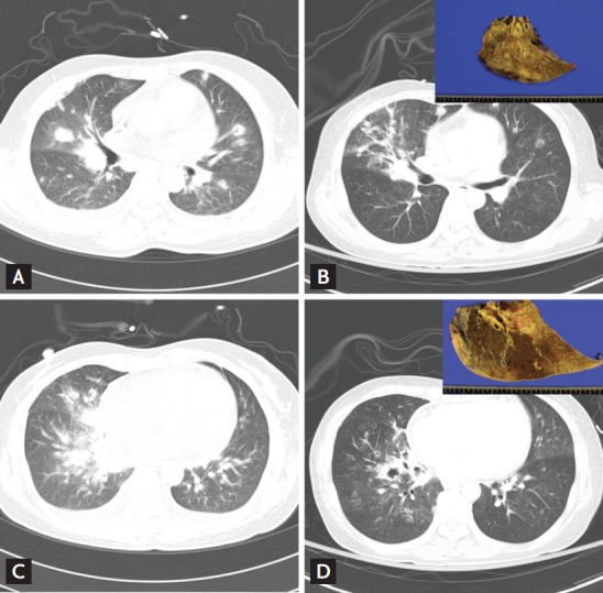
Figure 3.

Computed tomography (CT) images obtained from a 50-year-old female with angio-invasive pulmonary aspergillosis under induction chemotherapy. (A, B) High-resolution CT lung images (1-mm-thick) obtained at the level of the right middle lobe. Ill-defined macronodules with halo signs are evident in both lungs. One week after discharge, the patient presented to the emergency department with massive hemoptysis. (C, D) Conventional CT images (5-mm-thick) revealed that the number and size of nodules had increased in both lungs (especially the right lung), and cavitary changes were evident. The patient underwent bilobectomy of the right middle and right lower lobes. The pathology of the resected lung revealed aspergillosis featuring pulmonary artery invasion (insets in B and D).
