Figure 1.

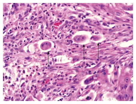
Representative histopathological micrograph of tumor budding (magnification × 400). Hematoxylin and eosin staining of a tumor section showing tumor budding (black arrows) at the invasive front.

Representative histopathological micrograph of tumor budding (magnification × 400). Hematoxylin and eosin staining of a tumor section showing tumor budding (black arrows) at the invasive front.