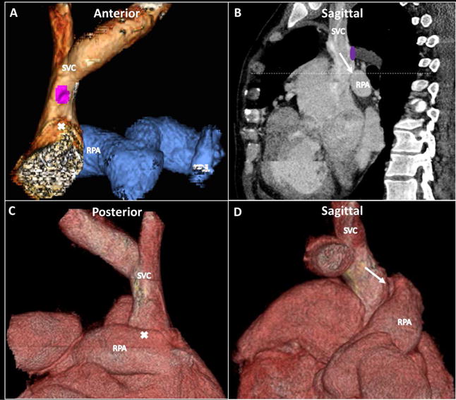
Figure 2. Contrast-enhanced Cardiac CT.

Pre-procedure contrast-enhanced computed tomography (CT) was used for procedure planning (A, C, and D: 3-dimensional (3D) reconstructions; B: sagittal plane) and included fluoroscopic visible landmarks (B, dashed line). CT imaging was used to plan target dimensions (anastomotic balloon expandable covered and anchoring bare-metal stent diameters) and understand distance requirements (covered stent lengths for anastomosis coverage without neighboring vessel occlusion). Purple circle = origin of azygous vein; white arrow = planned point and angle of exit and entry; white X corresponds to white arrow tip on corresponding alternate views. RPA = right pulmonary artery; SVC = superior vena cava.
