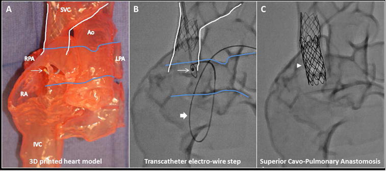
Figure 3. 3D-printed Heart Model.

(A) In this 3D-printed heart model based on the contrast-enhanced CT (view from anterior), the SVC is outlined in white and pulmonary artery in blue. The anchoring SVC stent is in place with the guiding catheter (arrow) positioned in the SVC prior to vessel traversal. Subsequent key procedural steps seen with x-ray fluoroscopy of the 3D-printed heart model show (B) the transcatheter electro-wire step with electrified 0.014” guidewire aimed from SVC (arrow) to RPA (loop snare catheter; block arrow); the snare catheter course was retrograde from aorta to single ventricle to right pulmonary artery; and (C) end-to-side anastomotic endograft (arrowhead) in place. Ao = aorta, IVC = inferior vena cava, LPA = left pulmonary artery, RA = right atrium, other abbreviations as in Figure 2.
