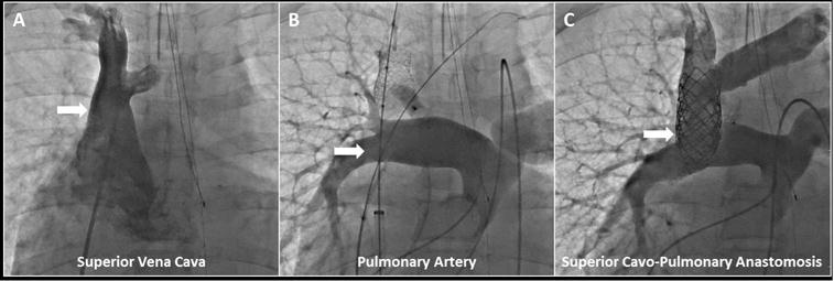
Central Illustration. Transcatheter Superior Cavopulmonary Anastomosis: Contrast X-ray Angiography.

(A) Pre-intervention superior vena cava angiogram was performed with the angiographic catheter introduced from femoral venous access, while (B) pre-intervention pulmonary artery angiogram was performed with the angiographic catheter introduced from femoral arterial access (retrograde to single ventricle through stenotic pulmonary valve). Superior vena cava anchor stent (and post-deployment balloon, sheath, wire), left innominate marking wire, and pacing catheter introduced from femoral venous access are shown. Angiographic catheter (introduced from right internal jugular) and deployed azygous vascular plug are also shown. (C) Post-transcatheter superior cavopulmonary anastomosis is seen via simultaneous angiography performed with previously described angiographic catheters introduced from femoral arterial and right internal jugular access.
