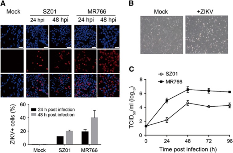
Figure 2.
ZIKV infection in the HK2 cell line. (A) Immunofluorescence staining of HK2 cells infected with SZ01 or MR766 ZIKV using an anti-ZIKV envelope protein antibody. Upper panel, representative images. Scale bar, 20 μm. Lower panel, quantification of infected cells. (B) Light microscopy images showing SZ01 ZIKV-induced CPEs. (C) Production of infectious SZ01 and MR766 ZIKV in HK2 cells. Cytopathogenic effects, CPEs.

