Figure 3.
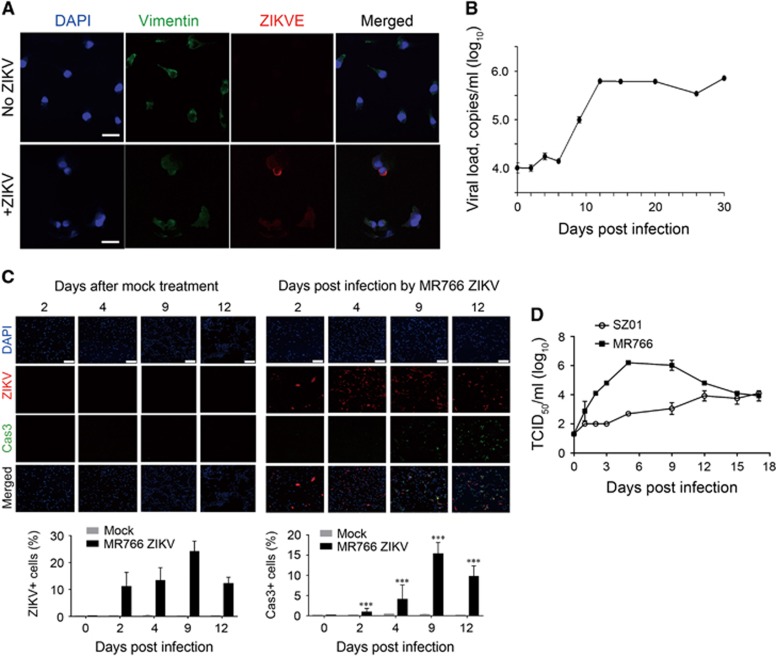
ZIKV infection in primary hRPTEpiCs. (A) Immunofluorescence staining of primary hRPTEpiCs infected with SZ01 ZIKV at a MOI of 2.5 for 96 h. Scale bar, 30 μm. (B) Long-term persistence of SZ01 ZIKV in primary hRPTEpiCs. Viral copies in the supernatant of medium were determined by RT-qPCR using ZIKV standard samples. Values are shown as the mean±standard deviation (sd; n=5). (C) MR766 ZIKV-induced apoptosis in primary hRPTEpiCs. Upper panel, representative immunofluorescence images. Scale bar, 200 μm. Lower panel, quantification of ZIKV-positive and caspase-3-positive cells. ***Significant difference between mock and infected cells (P<0.001; Student’s t-test). (D) Production of infectious SZ01 and MR766 ZIKV particles in primary hRPTEpiCs.

