
Figure 2.
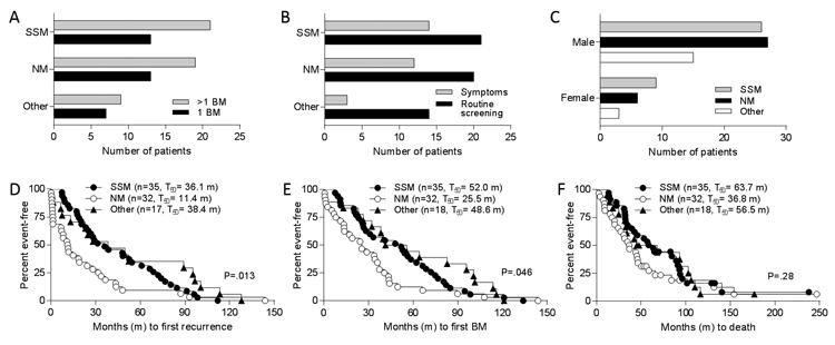
Analysis of primary tumor histologic pattern. Distribution of superficial spreading (SSM) and nodular (NM) melanoma types among patients A, with numerous (>1) or solitary (1) brain metastases (BM) detected; B, based on detection setting for BM (symptoms vs. routine screening); and C, by patient sex. Two-sided Fisher's Exact tests were used for analyses of primary tumor histologic pattern and number of BM, detection setting, and sex (see text). Patients with BM were grouped by primary tumor histologic pattern and times (in months, m) to D, development of first recurrence; E, diagnosis of first BM; and F, death were plotted. Number of patients (n) in each group and median times to events are shown in parentheses. P values based on log rank tests.