Figure 1.
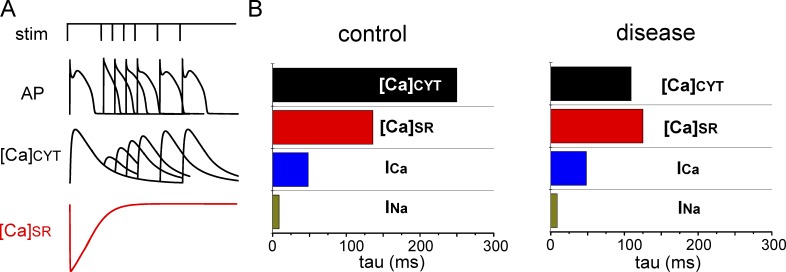
Shortened Ca-signaling refractoriness in cardiac disease. (A) Schematic illustration of a two-pulse protocol experiment showing the recovery of electrically stimulated Ca release is significantly delayed in relation to the recovery of electrical excitability. (B) Exponential constants (tau) of the recovery of Ca-transient amplitude ([Ca]cyt), free SR Ca ([Ca]SR), the amplitude of L-type Ca current (ICa), and peak of voltage-gated Na current (INa), are shown for control and diseased myocytes. Data for [Ca]cyt and [Ca]SR were obtained from the two-pulse experiments in ventricular myocytes from control and diseased hearts (Belevych et al., 2011, 2012). Rates of recovery for INa and INa were obtained with the Hund and Rudy (2004) model of the canine ventricular myocyte. Note the shortened time delay between the recovery of [Ca]cyt and [Ca]SR in diseased hearts.

