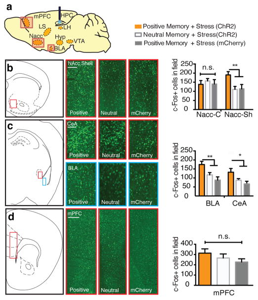
Figure 2.
Positive memory reactivation increases c-Fos expression in the nucleus accumbens shell and the amygdala. a, Brain diagram illustrating target areas analyzed. b, Activation of a positive memory, but not a neutral memory or mCherry only, in the DG during the TST elicits robust c-Fos expression in the nucleus accumbens shell (b), basolateral amygdala, and central amygdala (c), but not in the medial prefrontal cortex (d). For histological data, a one-way ANOVA followed by a Bonferroni post-hoc test revealed a significant increase of c-Fos expression in the positive memory + stress group relative to controls in the NAcc and amygdala, but not the mPFC (NAcc Shell: F2, 30 = 15.2, P < 0.01; BLA: F2, 30 = 11.71, P < 0.01; CeA: F2, 30 = 11.45, P < 0.05; mPFC: F2, 30 = 1.33, P = 0.294. n = 6 per group, 3–5 slices per animal). n.s., not significant, *P<0.05, **P<0.01,***P < 0.001. Data are means +/− SEM. Scale bars correspond to 100μm.

