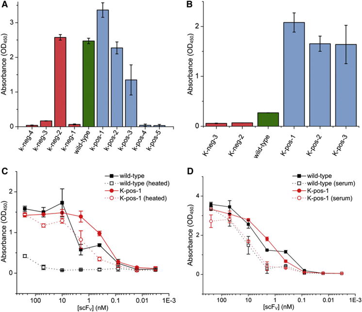
Figure 3. ELISA Screening and Characterization.

(A) The initial screen of the supercharged scFV variants for binding. ScFVs were added to the antigen-bound plate at a concentration of 5 μg/ml.
(B) Samples of constructs shown to bind in the initial screen were stored at 70°C for 1 hr, allowed to cool to room temperature, and assayed again to determine which constructs retained activity.
(C) A dilution-series ELISA comparing wild-type and K-pos-1 binding before and after incubation at 70°C for 1 hr.
(D) A dilution-series ELISA comparing the function of wild-type and K-pos-1 in 50% serum. Error bars represent SD calculated from the OD450 values of three identically-prepared wells.
See also Figures S2 and S3.
