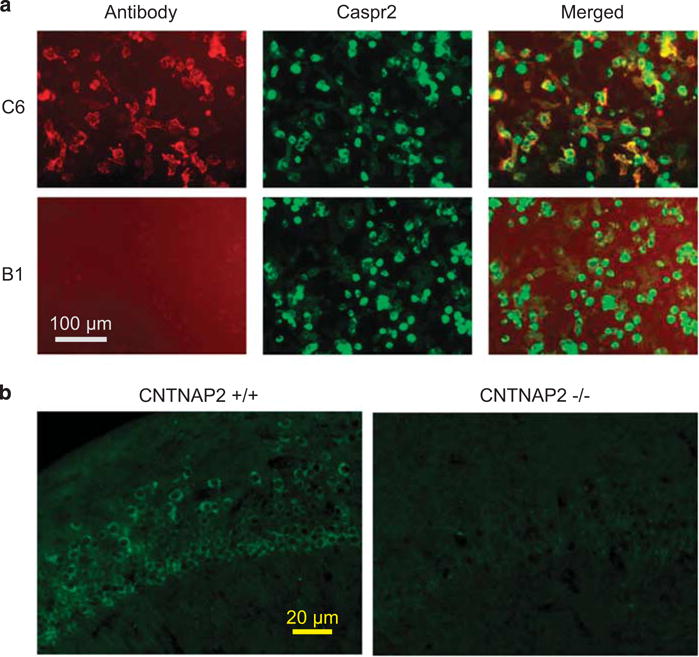
Figure 1.

Brain-reactive monoclonal antibody C6 binds to Caspr2. (a) C6 (top panels), but not control B1 (bottom panels) antibody co-localize with Caspr2 on HEK-293T cells, expressing tGFP-Caspr2. No staining was seen on cells expressing only tGFP or non-transfected cells (data not shown). (b) C6 antibodies show reduced staining to the CA1 region in the hippocampus of CNTNAP2−/− (right) compared with wild-type (left) mice. Caspr2, contactin-associated protein-like 2; tGFP, turbo-green florescent protein.
