Fig. 3.

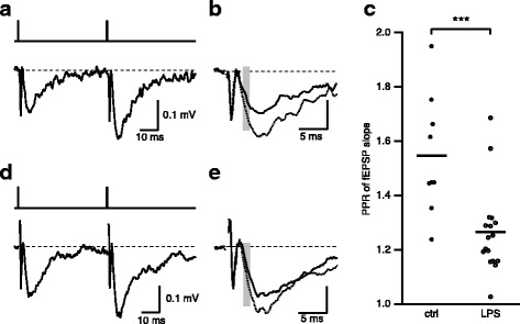
LPS exposure in utero causes reduced PPR. a, b Representative fEPSPs evoked by paired stimulation separated by 50 ms (top trace) from hippocampal slices obtained from adult control mice (a) and mice exposed to LPS in utero (d). Each trace is the average of 10 consecutively recorded voltage traces. b, e Overlay of the first (black) and second (dash line) fEPSPs. Gray bars highlight the regions where fEPSP initial slopes were measured. c Scatterplot of PPR determined by measuring the slope of the average of 10 individual trials. Asterisks indicate p < 0.001
