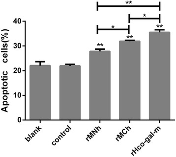
Fig. 6.

Apoptosis analysis of PBMC in response to rMNh, rMCh, and full-length Hco-gal-m by flow cytometry. Flow cytometric analysis of PBMC treated with recombinant proteins or recombinant empty protein pET-32a (control). Apoptosis of PBMC was determined by staining with annexin V and PI. The percentages of cells with different staining patterns are shown. The apoptosis rate was calculated by the percentage of early (AnnexinV + PI-) and late (AnnexinV + PI+) apoptotic PBMC. The percentage of apoptosis was measured on four separate occasions. PBMC used for all replicates of distinct treatments in each experimental repetition were derived from the same goat. Results presented here are representative of three independent experiments. Data are presented as the mean ± SD, **P < 0.001 vs the control group, a capped line designates two groups that differ significantly (*P < 0.01, **P < 0.001)
