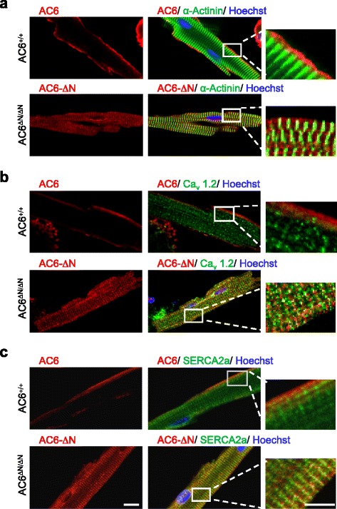
Fig. 3.

Cellular localization of AC6 and flag- AC6-ΔN in cardiomyocytes. Localization of AC6 (red) or flag- AC6-ΔN (red), and α-actinin (a, green), Cav1.2 (b, green), and SERCA2a (c, green) in adult cardiomyocytes was assessed by immunofluorescence staining. Scale bar, 10 μm. The rightmost panels show the enlarged, merged images of fields in white rectangles. Scale bar, 5 μm
