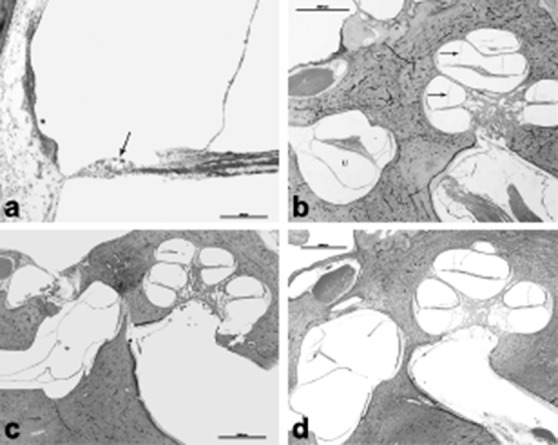
Fig. 3.

a) HB 655 Lt 130 10x: A higher magnification of the cochlea showed outer hair cell loss (arrow) and stria vascularis atrophy (*), b) HB 655 Lt 100 1x: 72-year-old male patient diagnosed with Ménière's disease. He had a history of bilateral fluctuating hearing loss, tinnitus and vertigo episodes. Histopathologic exam of the left ear showed profound cochlear hydrops (arrow). Utricle (U), c) HB650Lt110. This 79-year-old patient had a history of bilateral profound mixed type hearing loss and left stapedectomy. She was extremely vertiginous right after the surgery for months. Histopathologic exam showed bilateral otosclerosis, which is located anterior to the oval window. This slide shows the surgery site, otosclerosis and hydropic saccular membrane that showed healed perforation. Interestingly, cochlea did not show any hydropic changes, d) HB593Lt 470. This was an 86-year-old patient who had a history of Ménière's disease and 8th nerve resection. Histopathologic exam showed profound hydropic changes in the cochlea, utricle and saccule. Note the outpouching, and hydropic saccular membrane.
