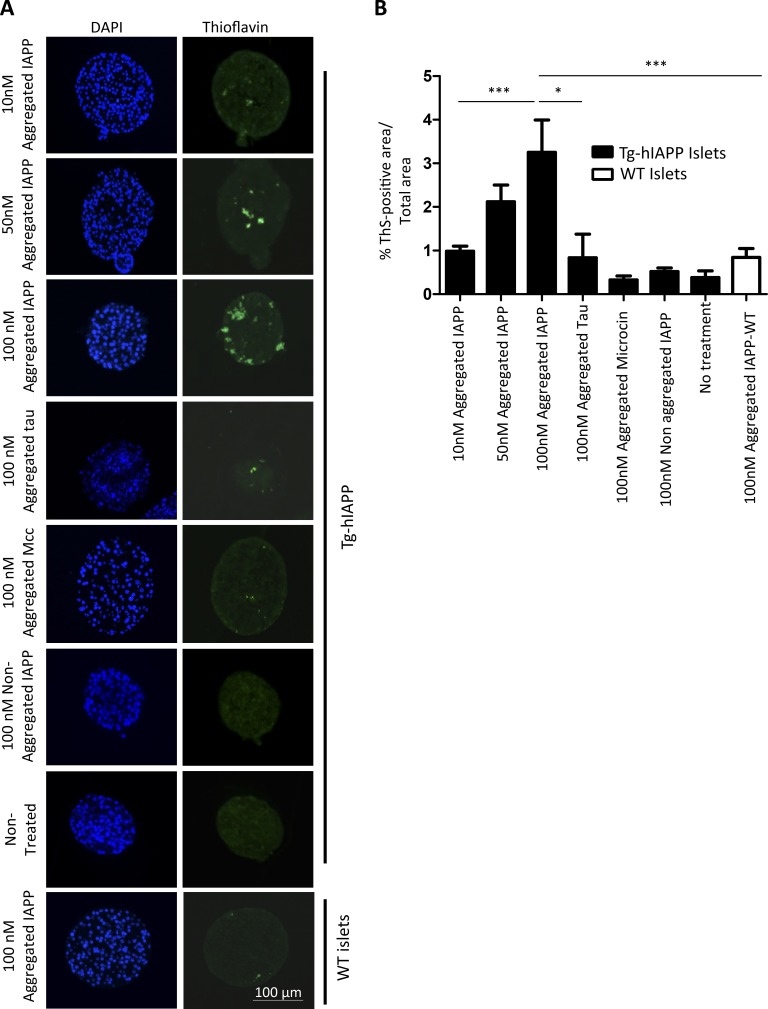
Figure 5.
Induction of IAPP amyloid deposition in islets by incubation with synthetic IAPP aggregates. (See also Figs. S3 and S4.) (A) Isolated islets from 3-wk-old, female, Tg-hIAPP mice were cultured in presence of different concentrations of IAPP aggregates prepared in vitro from synthetic IAPP, as well as controls treated with other amyloidogenic proteins, including the Alzheimer's disease–associated protein Tau (the K18 fragment) and the bacterial amyloid Mcc. Representative images of islets after diverse treatments, characterized by DAPI staining (blue) and the presence of amyloid by ThS amyloid staining (green). (B) The amyloid load present in the islets was quantified by measuring ThS-positive area/total islet area. The values correspond to means ± SE of 4–27 islets analyzed per condition. Data were analyzed by one-way ANOVA, and differences were highly significant (P < 0.0001). Individual differences among the different groups were studied by the Tukey’s multiple comparison test. *, P < 0.05; ***, P < 0.001.

