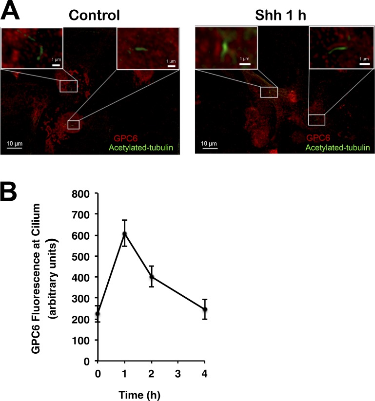
Figure 10.
Shh induces the migration of GPC6 in primary chondrocytes. (A) Chondrocytes isolated from E18.5 femurs were treated with Shh- or vector control–conditioned medium for 1 h. Then, cells were immunostained with antibodies for GPC6 (red) and acetylated tubulin (green). Boxes in the corners represent shifted overlays of enlargements of the indicated areas containing the cilium (bars, 1 µm). (B) GPC6 fluorescence at the cilia of chondrocytes was measured before and after Shh treatment for the indicated time points. The fluorescence intensities shown represent the mean of measurements performed in the following number of cells: 28 (0 h), 27 (1 h), 12 (2 h), and 12 (4 h) ± SD.

