Figure 6.
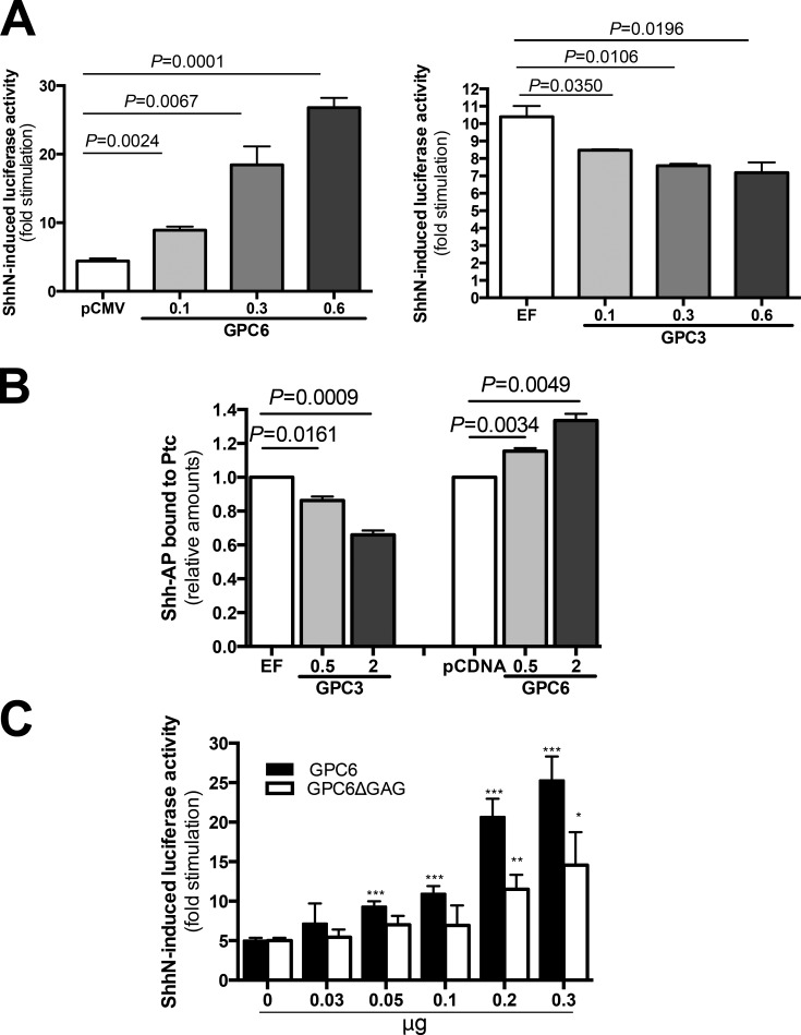
GPC6 stimulates the Hh signaling pathway. (A) Hh reporter assay. NIH3T3 cells were transfected with GPC6 (left) or GPC3 (right) or vector control (cytomegalovirus promoter [pCMV] and elongation factor promoter [EF]) along with a luciferase reporter driven by an Hh-responsive promoter and β-galactosidase. Transfected cells were stimulated during 48 h with ShhN- or control-conditioned medium. Then, luciferase and β-galactosidase assays were performed. Bars represent luciferase activity fold stimulation induced by Hh (mean + SEM of triplicates) normalized by β-galactosidase activity. One representative experiment of four is shown. (B) Increased binding of Shh to Ptc1 in the presence of GPC6. NIH3T3 cells expressing increasing amounts of GPC3 or GPC6 were incubated for 2.5 h at 4°C with Shh-AP or AP. Then, Ptc1 was immunoprecipitated, and the amount of Shh bound to the immunoprecipitated Ptc1 was determined by measuring AP activity. Bars represent the relative amount of Shh-AP bound to Ptc1 (mean + SEM of four independent experiments) after subtraction of the binding measured for AP alone. Shh-AP binding to the cells transfected with vector control was arbitrarily considered 1. (C) HS chains are required for maximum GPC6-induced stimulation of Hh signaling. NIH3T3 cells were transfected with increasing amounts of the indicated expression vectors along with a luciferase reporter driven by an Hh-responsive promoter and β-galactosidase. Then, luciferase activity in the presence and absence of Shh was performed as described in A. Bars represent luciferase activity fold stimulation induced by Hh (mean + SEM of triplicates) normalized by β-galactosidase activity. One representative experiment of two is shown. *, P < 0.05; **, P < 0.005; ***, P < 0.001. Statistical analysis was performed by Student’s t test (unpaired two-tailed).

