Figure 5.
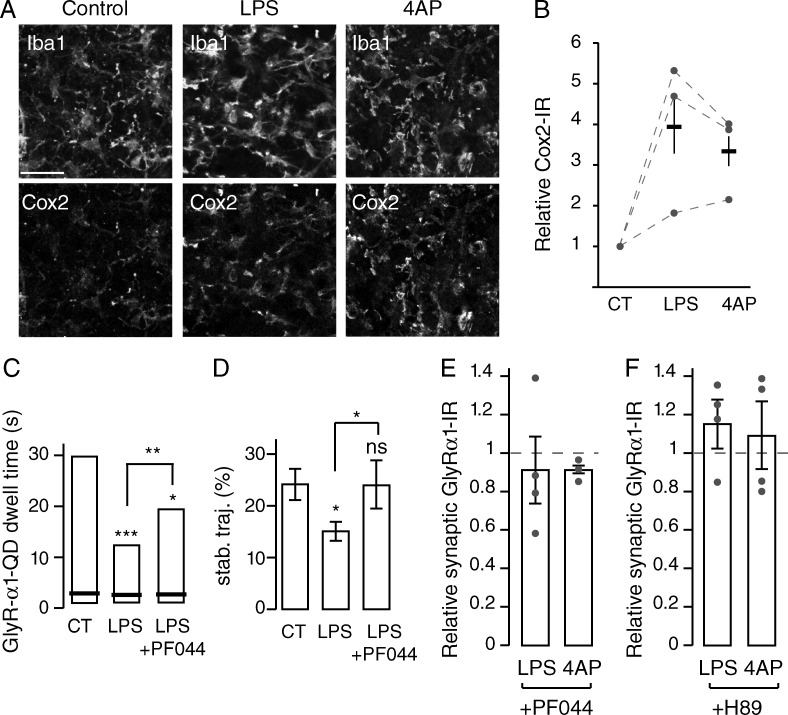
PGE2 mediates microglial regulation of synaptic GlyR accumulation via EP2 receptors. (A) Double labeling of organotypic slices showing Cox-2 and Iba1 IRs in control condition and after 30-min LPS application or 15-min 4AP application. Cox-2 IR is restricted to microglia. Bar, 20 µm. (B) Quantification (mean ± SEM) of Cox-2 fluorescence intensities over the corresponding Iba1 profiles in control (CT), LPS, and 4AP conditions. Circles represent single experiments. (C and D) The modulation of GlyR α1 QD synaptic dwell time (C) and stability (D) in control (CT), after LPS application (LPS), and after LPS and PF04418948 treatment (LPS + PF044). (C) Synaptic dwell time distributions indicating 25, 50 (black bar), and 75% of all trajectories. *, P < 0.05; **, P < 0.01; ***, P < 0.001; Kolmogorov-Smirnov test. (D) Percentage of stable synaptic trajectories detected during the imaging session (n = 3 independent experiments; mean ± SEM; ns, P > 0.05; *, P < 0.05; t test). (E and F) Fluorescence intensities relative to control of synaptic GlyR α1 IR after application of LPS or 4AP in the presence of EP2 receptor antagonist PF04418948 (E) or the PKA antagonist H-89 (F) in organotypic slices. Mean ± SEM; circles represent single experiments.

