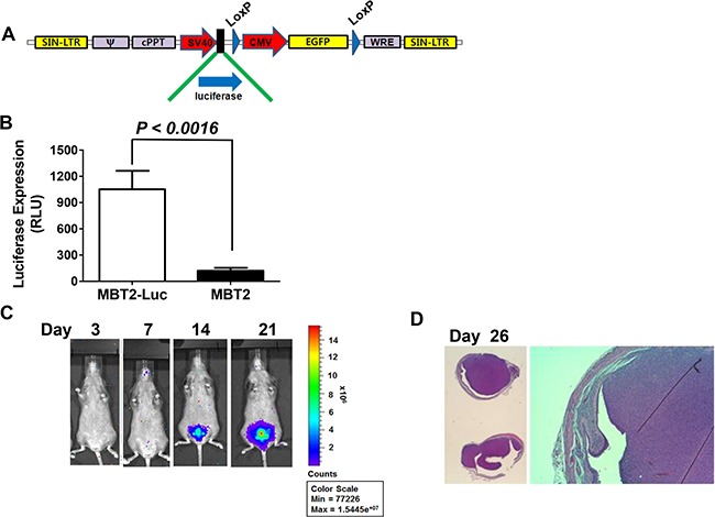
Figure 1. Construction of MBT2.Luc cells expressing the luciferase gene.

(A) The lentivirus shuttle vector pLoxLL3.7 was engineered to express a luciferase expression cassette under the SV40 promoter. Then, a four-plasmid-based lentiviral expression system was co-transfected with pMDLg/pRRE, pRSV-Rev and pCMV-G. (B) Approximately 1 × 10⁵ MBT2.Luc and MBT-2 cells were prepared in 12-well plates for 24 h. Cells were harvested for luminescence measurements as described in the Materials and Methods. The data represent the mean ± SD. (C) To establish the orthotopic bladder cancer model, 50 μl of 0.1 μg/mL poly-L-lysine was instilled for 15 minutes, and the bladder was voided. Approximately 2 × 10⁶ cells suspended in PBS were instilled intravesically via the urethra using a 22-gauge arterial puncture needle cannula. Luminescence images were captured at the indicated time points using INVIVO Lumina. (D) For histological analyses, all tumors from five mice were harvested at day 26. Tumor sections (5–10-mm thick) were affixed to slides, de-waxed with ethanol, and stained with hematoxylin and eosin (H&E).
