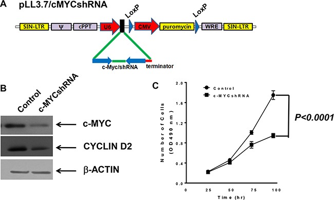
Figure 3. MBT2.cMYCshRNA cells expressing shRNA showed reduced numbers of c-myc transcripts.

(A) The lentivirus shuttle vector pLoxLL3.7 was engineered to express c-myc shRNA under the U6 promoter (pLL3.7/cMYCshRNA). Then, a four-plasmid-based lentiviral expression system was co-transfected with pMDLg/pRRE, pRSV-Rev and pCMV-G. (B) Cell lysates of MBT-2 and MBT2.cMYCshRNA were prepared and resolved on SDS-PAGE for western blotting. Western blotting using c-MYC-reactive or cyclin D2 antibodies showed the reduction of c-myc mRNA and the down-regulation of the cyclin D2 gene. (C) Approximately 3 × 10³ cells of each cell line were plated in a 96-well plate for 48 h, and proliferation was evaluated using MTT assays. P values were determined by two-tailed paired t-tests.
