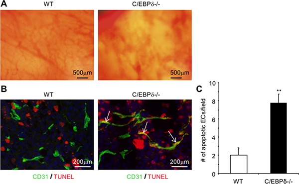
Figure 4. Loss of C/EBP-δ results in increased endothelial apoptosis and hemorrhagic vascular morphology in tumors.

Dorsal skin vascular window chambers were established in WT and C/EBP-δ null mice, and 1 × 10⁵ 3LL tumor cells were injected into each window chamber. Tumor angiogenesis was imaged in live mice under microscopy 10 days after tumor cell implantation (Panel A). Tumor were harvested, sectioned and co-stained with an anti CD31 antibody (green) and TUNEL assay (red) (Panel B). The number of double positive cells was counted in 10 randomly selected high power fields under microscopy (Panel C). Representative images are shown. n = 5 mice per group. **p < 0.01.
