Figure 2.
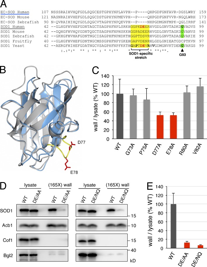
Identification of diacidic residues for SOD1 export. (A) Alignment of partial amino acid sequences of human, mouse, and zebrafish EC-SOD, and human, mouse, zebrafish, fruit fly, and budding yeast SOD1. The SOD1-specific stretch is shown in yellow, the residues in yeast SOD1 mutated to alanine in this study are in bold, the diacidic DE77/78 motif in human and yeast SOD1 are in bold red letters, and the G93 residue in human SOD1 mutated to alanine in ALS and its equivalent in yeast SOD1 are shown in bold and highlighted in green. Asterisks indicate positions that have a single fully conserved residue, colons indicate conservation between groups of strongly similar properties, and periods indicate conservation between groups of weakly similar properties. (B) Superimposition of the 3D structures of the human SOD1 monomer (PDB code: 2C9U) and human EC-SOD monomer (PDB code: 2JLP). SOD1 and EC-SOD are shown in gray and blue, respectively; the SOD1-specific amino acid stretch is shown in yellow, and side chains of D77 and E78 are in red. (C) Effect of single-alanine substitutions in the SOD1-specific stretch on yeast SOD1 secretion. Cells expressing either wild-type yeast SOD1 or single alanine substitutions in positions G73, P75, D77, E78, R80, or V82 of yeast SOD1 were grown to mid-logarithmic phase, washed twice, and incubated in 2% potassium acetate for 2.5 h. Cell wall proteins were extracted from equal numbers of cells followed by precipitation with TCA. Lysates and cell wall–extracted proteins were analyzed by Western blot. The ratio of wall to lysate SOD1 was determined and represented as percentages of the strain expressing wild-type SOD1. (D) The DE77/78 diacidic motif is essential for yeast SOD1 secretion. Representative cell wall extractions monitored by Western blotting of cells expressing either wild-type, DE77/78AA, or DE77/78NQ yeast SOD1 incubated for 2.5 h in 2% potassium acetate. (E) SOD1 levels were quantified, and the ratio of wall to lysate SOD1 was determined and represented as percentages of the starved wild-type culture. (165X) wall indicates that the wall sample represents the material from 165-fold the number of cells analyzed in the lysate sample. Results in C and E are shown as means ± SEM of at least three experiments.

