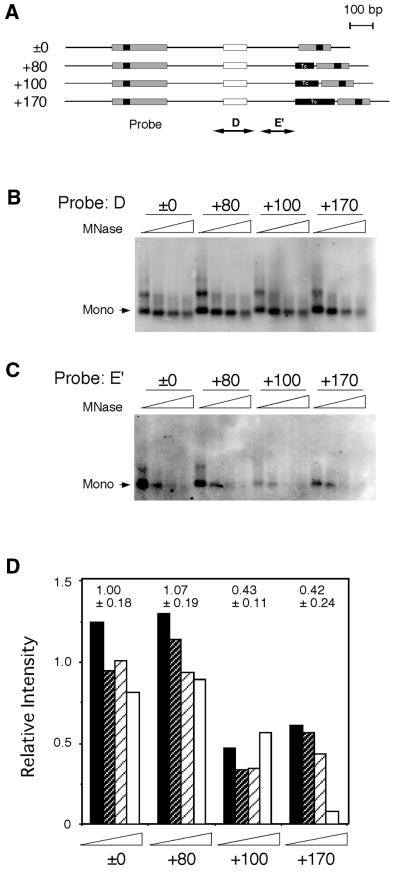
Figure 4.
In vitro nucleosome phases at HS2. (A) Constructs with various distances between ɛB-16 and NF-E2 sites were reconstituted with core particles in vitro. Reconstituted nucleosomes were digested with micrococcal nuclease, and DNA fragments were purified and subjected to Southern blot analysis. The probes corresponded to the nucleosome phase of the NF-E2 region (probe D) and the next phase toward ɛB-16 (probe E′). The same membrane was stripped and used for rehybridization. (B) Southern blot analysis of nucleosomes at NF-E2 site. (C) Southern blot analysis of nucleosomes in the region E′. (D) Relative degrees of nucleosome formation at region E′. The intensity of the bands was quantified by ImageQuant (Molecular Dynamics), and the degrees of nucleosome formation at region E′ relative to those of NF-E2 (determined with probe D) were calculated. The averages and the standard deviations, which were normalized with the average value for ±0 construct, are shown above.

